Disclaimer
All content provided on this blog is representation of the blog owner and not Franciscan University of Steubenville. The information on this site is purely used for education purpose. The owner of this blog makes no representations as to the accuracy or completeness of any information on this site or found by following any link on this site. The owner will not be liable for any errors or omissions in this information nor for the availability of this information. The owner will not be liable for any losses, injuries, or damages from the display or use of this information.
Privacy
The owner of this blog does not share personal information with third-parties nor does the owner store information is collected about your visit for use other than to analyze content performance through the use of cookies, which you can turn off at anytime by modifying your Internet browser’s settings. The owner is not responsible for the republishing of the content found on this blog on other Web sites or media without permission.
Blog Comments
The owner of this blog reserves the right to edit or delete any comments submitted to this blog without notice due to;
1. Comments deemed to be spam or questionable spam
2. Comments including profanity
3. Comments containing language or concepts that could be deemed offensive
4. Comments that attack a person individually
This policy is subject to change at anytime.

Thursday, October 17, 2013

Endospore Stain

Next we wanted to see if our bacteria possessed any endospores. An endospore is a mechanism used by bacteria that is specialized for the survival of that bacteria in unfavorable conditions.

We began this staining procedure much like we did for the Acid Fast stain. First we smeared some of our sample bacteria on the the slide and using a Bunsen burner we heat fixed the bacteria to the slide so that it can we stained and viewed under a microscope.
After that we put a piece of bibulous paper over the slide and using forceps, moved the slide onto a drying rack over a beaker of boiling water.


Next we saturated the paper with malachite green solution. Just like the acid fast stain we had to remain attentive and continue to add drops so that the solution did not evaporate and dry out the slide. 
We continued this practice for 6 minutes, constantly adding more drops because the solution was evaporating very quickly from the bibulous paper. 


Using forceps we removed the bibulous paper from the slide and placed it in a bio-hazard bag. We then transported our slide back to our lab bench, using forceps, and placed it on a drying rack over the sink in order to let it cool off. We then rinsed the slide off with water to remove excess malachite green solution. 


 Next we covered the smear with safranin for 90 seconds. Then we washed off the excess safranin stain with water.  


To completely dry the stain we blotted the side between bibulous paper, being careful to not blot it too excessively.

Now our slide is ready to view under the oil immersion lens of our microscope. Upon viewing our bacteria we were able to conclude that our bacteria does not possess endospores. If our bacteria did we would be able to see little green dots at the ends of our bacteria rods. But our bacteria is completely red, so they do not have endospores. 


Acid Fast Stain

In order to find out if our bacteria is acid fast or non-acid fast we had to do an acid fast stain.
We first smeared some of our bacteria on a slide and heat fixed it to the slide (just as we have been doing for all our stains) so the bacteria will not be washed off by all of the liquids it will be encountering. Once the slide was heat fixed we put a square of bibulous paper on the slide and then put the slide on a drying rack that was resting over a beaker filled with boiling water.
Transporting our slide was really tricky because our forceps didn't hold onto our slide completely, so we were very careful in transporting our slide and very anxious that we might drop it and have to start over again!


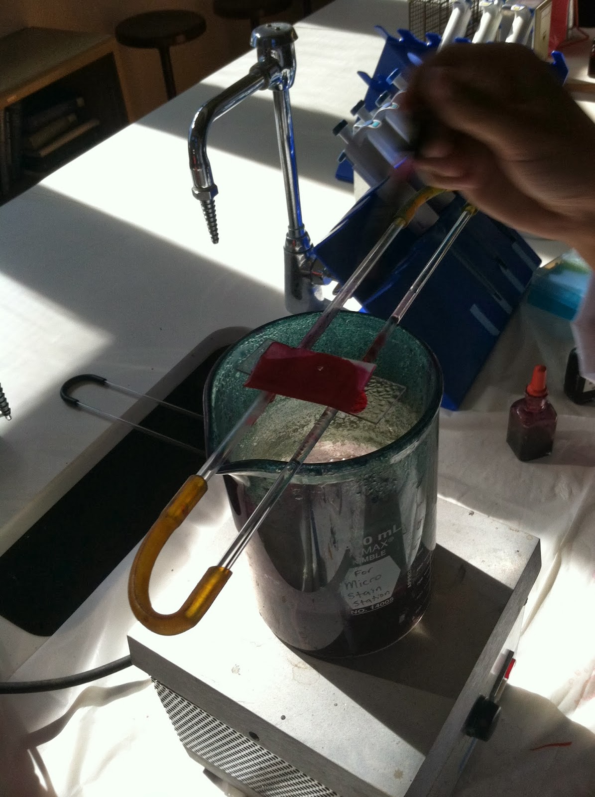
Then we proceeded to drop Kinyoun's carbolfuchsin solution over the slide for 5 minutes, all the while making sure the slide doesn't ever get too dry. 

We were sure to constantly drop the solution on the paper (so it would look like this) to make sure that it never dried out.

Next, with the slide still over the beaker of boiling water, we added Ziehl-Neelsen carbolfuchisn. We used the same method for this stain. For three minutes we watched and added more drops to our slide in order to make sure the solution wouldn't evaporate and dry out the slide. 

Using forceps we very carefully removed our slide from over the beaker of boiling water. (Which proved to be quite difficult for us, but in the end we were able to do it) We then placed the slide on a drying rack and removed the bibulous paper, disposing of it in a bio hazard bag. After letting the slide cool for a few minutes, we rinsed the slide off with water to remove any access stain. 
Then we proceeded to use a decolorizing agent, holding the slide at a 45 degree angle we dropped the agent on the slide until the color stopped running. After we washed off the slide with de-ionized water to remove the decolorizing agent. 

Next we covered the slide with methylene blue for 2 minutes, flooding the slide with it and then letting it sit. 


After 2 minutes we rinsed off the excess stain with water. To remove the water we then put our slide between two pieced of bibulous paper and gently blotted it to remove any water, but not too excessively so we would not ruin the stain we had made.

Now it is time to observe what we obtained by looking at our stained bacteria under the microscope, using the oil immersion lens!


Upon observing our bacteria, we could clearly see that all the bacteria had been stained blue, and retained none of the red staining. We concluded from this that our bacteria is in fact no-acid fast. 


Friday, September 27, 2013

Preparing a Capsule Stain

After doing a negative stain we now are going to do a capsule stain. By doing a capsule stain we are able to see if our bacteria have capsules or slime layers.

We first prepared a smear of bacteria in the Nigrosin stain.


Then after the smear is completely dry we covered it with the Safranin stain for one minute as indicated in our lab book.


After one minute, we gently washed off the excess stain. We had to make sure to avoid removing much of the stain. 


We, then, blot water from the slide with bibulous paper.


We now can see whether or not our bacteria from sample S have capsules or not! 


As you can see under the oil immersion lens of a microscope our bacteria do not have capsules because there is no space between the bacteria itself and the surrounding stain.


Preparing a Negative Stain of Sample S!

In lab we did a negative stain of sample S so that we could view the bacteria cells that are difficult to stain with basic dyes and are easily distorted by heat-fixation. Therefore, we created a dark background for the bacteria cells

To create this negative stain we first placed a tiny drop of Nigrosin stain near one end of a clean slide.


Then we used a sterilized inoculating loop to transfer a small amount of bacteria, taken from our sample S, into the Nigrosin stain drop.

 
We then mixed the bacteria into Nigrosin stain drop.


We made sure to flame our loop before we set it down and we grabbed a second microscope slide and touched the Nigrosin stain drop at a 30-45 degree angle and quickly pushed the slide to spread out the drop.

Notice how the slide now has a thin film containing the Nigrosin stain and the bacteria from sample S.


We then waited for the slide to air dry completely and once it was done air drying we examined the stained smear under the microscope using the oil immersion lens.

Look at what we saw!!


(Fun note: It looks like stars in the night sky. The bacteria represent the stars and the purple background represents the night sky. It looks really cool!)

A Closer Look at Sample S!!

When we removed our stock bacteria of sample S from the incubator we found the bacteria had grown in the same pattern as our first slanted agar plate and looked the same, so we were able to successfully transfer the bacteria.
Next we wanted to examine the bacteria more closely so put it on a microscope slide. Using the inoculating loop we got a bead of bacteria from our working culture (not the stock culture) and mixed it on the slide with a small drop of distilled water.
We waited for the slide to dry and then passed our slide through the flame of the Bunsen burner three times to heat fix our smear to the slide.

After this we proceeded to do a gram stain on our slide. We cover the slide with crystal violet for 20 seconds, then rinsed it off.

Next we covered the smear with gram's iodine for one minute, then rinsed it off.

Holding the slide at a 45 degree angle we dropped EtOH on the slide until the color from the stains stopped running. Immediately after we rinsed the slide and then covered the slide with safranin for one minute, and rinsed off the excess stain after.

To completely dry the slide we blotted it with bibulous paper, and then it was ready to be observed under the microscope. We put oil on the slide so that we could view the bacteria using the oil immersion lens. The oil is necessary because it has a refractive index that is the same as glass. Light can bend in air, so with the oil there, less light is lost and one is able to see their specimen more clearly.
By using the oil immersion lens we were able to see our sample more clearly, and this is what we observed!


 We found that our culture S was gram negative and the shape was long skinny rods. If you look closely you can even see some in the midst of binary fission. (they are the ones that look like long chains because they are multiplying to be many short rods)