Disclaimer
All content provided on this blog is representation of the blog owner and not Franciscan University of Steubenville. The information on this site is purely used for education purpose. The owner of this blog makes no representations as to the accuracy or completeness of any information on this site or found by following any link on this site. The owner will not be liable for any errors or omissions in this information nor for the availability of this information. The owner will not be liable for any losses, injuries, or damages from the display or use of this information.
Privacy
The owner of this blog does not share personal information with third-parties nor does the owner store information is collected about your visit for use other than to analyze content performance through the use of cookies, which you can turn off at anytime by modifying your Internet browser’s settings. The owner is not responsible for the republishing of the content found on this blog on other Web sites or media without permission.
Blog Comments
The owner of this blog reserves the right to edit or delete any comments submitted to this blog without notice due to;
1. Comments deemed to be spam or questionable spam
2. Comments including profanity
3. Comments containing language or concepts that could be deemed offensive
4. Comments that attack a person individually
This policy is subject to change at anytime.

Thursday, October 31, 2013

Indole (Tryptophan Degradation) Test

In the Indole test, we can determine the ability of our bacteria to split the amino acid tryptophan into indole and pyruvic acid.

We first used the aseptic technique to inoculate the tryptone broth tube with a loopful of bacteria from our agar slant culture. 


Then we incubated the inoculated tube at 35 degrees Celsius for 2 days. 

When we came back after two days we added 5 drops of Kovac's reagent to the inoculated tube.


As you can see there is a darker yellow that has formed on top of the lighter yellow broth. This means that the test came out negative. Therefore, our bacteria are not able to split the amino acid tryptophan into indole and pyruvic acid.


Nitrate Reduction Test

The Nitrate Reduction test determines if our bacteria is able to reduce nitrate ions to either nitrite ions or nitrogen gas.

We first obtained a nitrate broth tube and used the aseptic technique to inoculate the nitrate broth tube with a loopful of bacteria from our agar slant culture.


We then incubated the inoculated tube at 35 degrees Celsius for 2 days.

After two days, we added 5 drops of reagent A (sulfanilic acid) and 5 drops of reagent B (dimethyl-alpha-naphthylamine) to our inoculated tube. We made sure to wear gloves when we added the nitrate reagents.


After one to two minutes, we did not see a color change to pink or red color which means that we now need to add a small amount of powdered zinc to the tube.


After about 10 minutes we saw a small color change to our broth. This means that our bacteria are not able to reduce nitrate ions to either nitrite ions or to nitrogen gas.

Methyl Red Test and Voges-Proskauer Test

The Methyl Red test determines the ability of some bacteria to ferment glucose due to mixed-acid fermentation. While the Voges-Proskauer test determines the ability of some bacteria to ferment glucose via butanediol fermentation.

First we used the aseptic technique to inoculate the MR-VP (methyl red/Voges-Proskauer) broth tube with a loopful of bacteria from our agar slant culture.


Then we incubated the inoculated tube at 35 degrees Celsius for two days. 

After two days we came back to lab and divided the MR-VP broth into two tubes so that we could use the broth to use for another test. For one of the tubes we added 6 drops of the methyl red to the tube. We then gently swirled the tube to mix the broth culture and the pH indicator

After we mixed the broth culture and the pH indicator we read the reaction immediately and we found that our test was positive.


Then we took the other tube of the MR-VP broth and added 15 drops of Barritt's reagent A (alpha-naphthol) and 5 drops of Barritt's reagnet B (KOH) to the tube.


We then tapped the bottom of the tube vigorously so that the oxygen in the air aerates the medium.

Afterwards we put the tube back in our rack for about 30 minutes. After 30 mines we read our results and found that our bacteria was positive which means that our bacteria used the butanediol fermentation pathway.


Litmus Milk Reactions

The litmus milk reactions allows us to see our bacteria's ability to utilize lactose, protein, and litmus in litmus milk. 

First we obtained a litmus milk tube and used the aseptic technique to inoculate the litmus milk tube with a loopful of bacteria from our agar slant culture.


We then incubated the inoculated tube at 35 degrees Celsius for two days.

When we came back to lab two days later we found that our tube showed acid curdling with litmus reduction. Unfortunately, we did not get a picture of it as we accidentally mixed the tube forming what is shown below.
 

Fat (Triglyceride) Hydrolysis Test

We used the fat hydrolysis test to determine whether our bacteria are able to digest a triglyceride, which is a type of lipid.

First we obtained a tributyrin agar plate and we used the aseptic technique to inoculate the tributyrin agar plate with a loopful of bacteria from our agar slant culture.


We then incubated the inoculated plate upside-down at 35 degrees Celsius for two days.

When we came back to lab two days later, we took out our plate from the incubator and examined the plate for a clear area around our bacteria growth (as indicated on the left). We did not see a clear area around our bacteria growth so this indicates that our bacteria are not able to digest a triglyceride.


Gelatin Hydrolysis Test

We used the gelatin hydrolysis test to determine whether our bacteria are able to digest gelatin.

First we obtained a nutrient gelatin deep tube and used the aseptic technique to inoculate the nutrient gelatin deep tube by stabbing the tube with an inoculating needle containing our bacteria from our agar slant culture.


We the incubated the inoculate tube at 35 degrees Celsius for two days.

When we came back two days later, we took our nutrient gelatin deep tube from the incubator and placed in the refrigerator for 15 minutes. After 15 minutes, we took out our nutrient gelatin deep tube from the refrigerator and determined that it was negative. This means that unfortunately our bacteria are not able to digest gelatin.


Casein Hydrolysis Test

We used the casein hydrolysis test to determine whether our bacteria are able to hydrolyze or digest casein, which is the major protein in milk.

First we obtained a skim milk agar plate and then we used the aseptic technique to inoculate the skim milk agar plate with a loopful of bacteria from our agar slant culture.


We then incubated the inoculated plate upside-down at 35 degrees Celsius for two days.

After two days, we unfortunately found out that our test (on the right) was negative because after examining the plate we did not find a clear zone around our bacterial growth, which would have indicated a positive test.

Starch Hydrolysis Test

We used the starch hydrolysis test to see if our bacteria are able to hydrolyze or digest starch.

We first obtained a starch agar plate and used the aseptic technique to inoculate the starch agar plate with a loopful of bacteria from our agar slant culture.


We then incubated the inoculated plate upside down at 35 degrees Celsius for two days.

After incubation we flooded the starch agar surface with Gram's iodine. After flooding the agar plate we waited 30 seconds to a minute to determine whether our bacteria are able to digest starch.


After a minute we determined that our bacteria are not able to digest starch.


Testing For Sucrose, Glucose and Lactose!

In order to test if our bacteria would react with sucrose, glucose or lactose, we used the aseptic technique and transferred a small inoculating loopful of bacteria into a solution of each of these. We started out be sterilizing our inoculating loop, then we removed a small bead of culture from our original sample S. We flamed the opening of the tube and then put it away. We then transferred that bead of bacteria to first the sucrose solution. We stirred around the loop for a little bit to ensure that the bacteria was introduced into this new solution. Then flaming the mouth of the tube and the inoculating loop we incubated the solution at 37 degrees centigrade.  

We repeated the procedure above for the glucose solution, using the aseptic technique, and then putting the tube in the incubator. 


Our last solution was Lactose and we repeated this same process using the aseptic technique and incubating it at 37 degrees. 


Next class we would review the results we obtained from these tests. 


We discovered from the results that our bacteria utilized glucose, because it changed from a pink color to yellow. 
It partially utilized sucrose, indicated by the orange-ish color, but not fully yellow yet. 
We could determine that our bacteria did not utilize lactose, because it did not change at all from its original pinkish color. 


Motility Testing

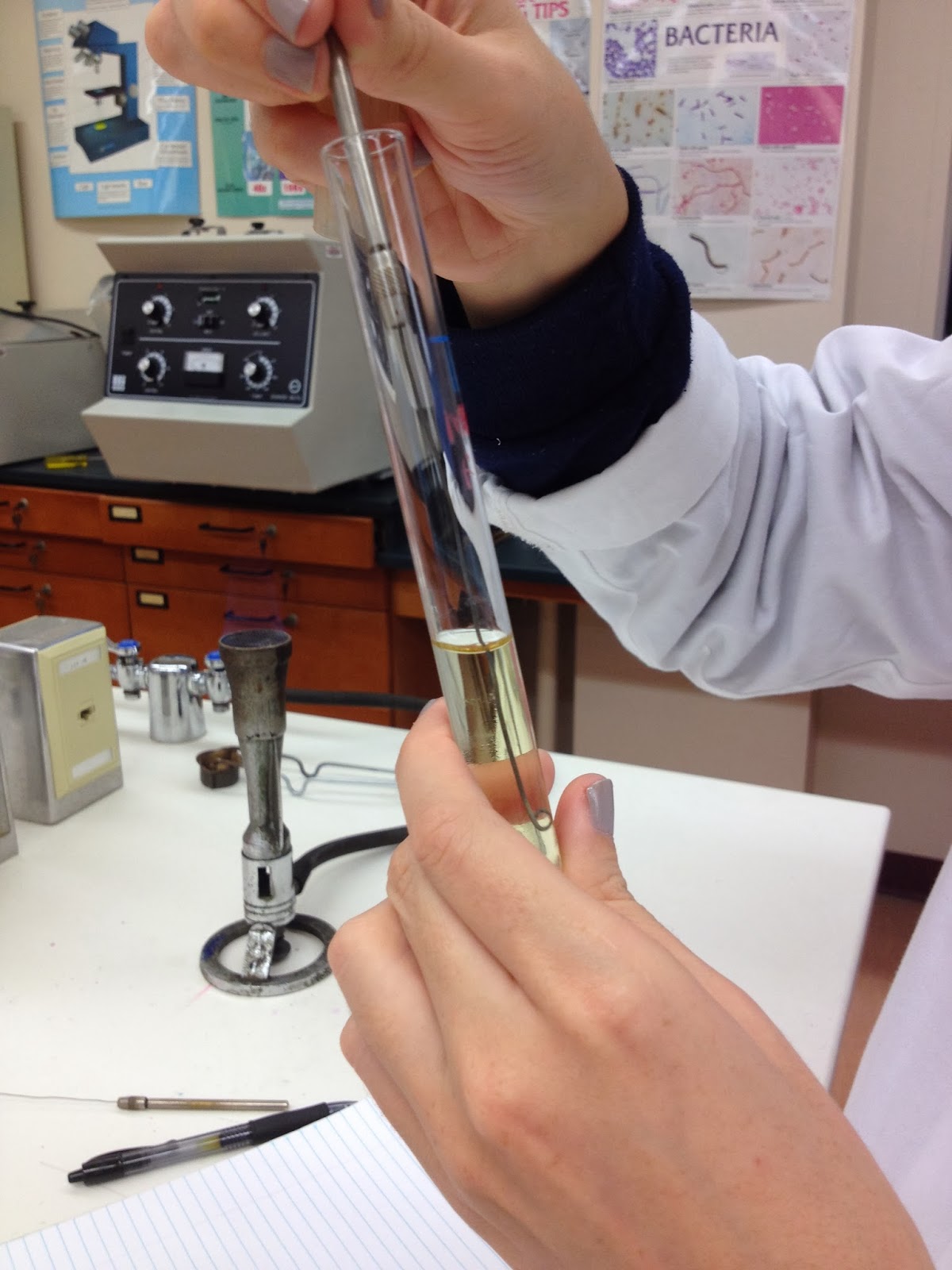
In lab we did a motility test to find out whether our sample S bacteria have one or more flagella that are propelling the bacteria.

First we have to obtain a sample of bacteria from our sample. We do this by touching the bacteria with a sterile inoculating needle. Then we stab the bacteria into the center of the test medium to about halfway to three-quarters depth.


We also did the same technique with a broth culture.


RESULTS:

After incubating our bacteria at 37 degrees centigrade, we were able to observe that our bacteria is motile!
The bacteria that was in the semisolid agar medium, not only grew along the stab of the innoculating needle, but also swam outward creating a small cloud of bacteria in the test tube. 


We can conclude from this test that our bacteria possesses some sort of flagella, that spins in a corkscrew motion and propels the bacteria making it motile.
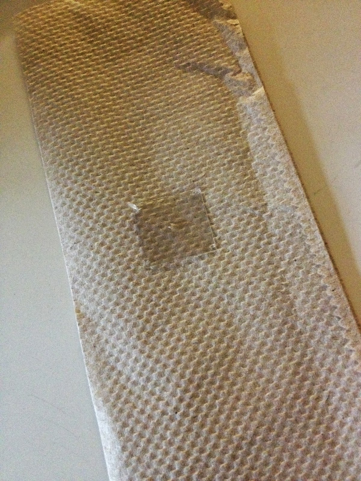
We further tested our bacteria's motility by observing the broth we incubated on a microscope slide. 
To do this we had to make a hanging drop slide. 
We first got a clean depression slide a coverslip and some petroleum jelly. 

On the corners of the coverslip we put four small dots of petroleum jelly.
 Then using a sterile innoculating loop we obtained a bead of our bacteria from the broth culture, and put that bead in the center of our coverslip. 


Next we gently put the cover slip on the depression slide. This resulting in the drop hanging over the depression in the slide so that bacteria could be viewed swimming within the drop.

This procedure proved to be very difficult for us, and it took us multiple tries to to be able to actually see the bacteria. When we finally did, we confirmed (with alot of help from Dr. P) that our bacteria was motile!
 We were able to see our rod shaped bacteria actually swimming around!!